
Cellometer Protocol
Nexcelom Cellometer Vision Putnam Lab Protocol
PROTOCOL IN PROGRESS
Cellometer Vision
Cellometer Vision located in Dr. Meenach’s lab in the URI Engineering building.
Cellometer Fluorescent Viability Cell Counters here. We use a Cellometer Vision CBA Image Cytometer (link here.)
Cell counting slides from Nexcelom: SD100 slides.
Equipment to transport to Engineering Building
- 20 uL pipette tips; 20 uL pipette (Either P20 or P200).
- Samples in a freezer bead box
- Gloves
- Trash bin (for pipette tips and slide protective covering)
- Cellometer slides
- mini vortex
- kimwipes
Protocol
- Turn on Nexcelom laptop. Turn on the cellometer by clicking the on/off switch in the back of the machine.
- Open the ‘Cellometer Vision CBA’ program.
- Select the ‘Putnam Lab Dinoflagellate Assay’ program under ‘Assay Type’ on the left hand ‘SETUP’ bar.
- Imaging mode will read ‘Flourescence Only’ and the cell count will come from F1 flourescence.
- Take a cellometer slide and remove the protective film from the bottom and then the top of the slide.
- Vortex the homogenate sample.
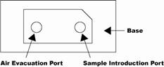
- Load the slide by pipetting 20 uL into the circle insert on one half of the slide. The sample is placed in the ‘Sample Introduction Port’ (see image below).
- Place slide directly into the sample input slot on the cellometer vision. The ‘Cellometer Vision CBA’ program will register that a slide is inputted.
- Select ‘Set User/Sample ID’. User= ‘Emma Strand’. Sample ID example = ‘1686-1’. Four digit coral ID number and then the replicate number. 1686-1 means coral #1686 and cell count replicate 1. Select ‘save’.
- Dilution factor = 1.
- Exposure = 3,000 msec.
- Select ‘Preview F1 Image’.
- Select ‘Count’. The machine will image four squares and output the cell counted value for those squares and a value converted to cells per mL.
- Record the cells counted and concentration values. You can also save and output these values as an excel spreadsheet at the end.
- Remove the slide and mark an ‘X’ in sharpie on the side that has been used (see image below).
- Repeat the above process 2 more times to get a total of 3 counts per sample.
- Clean the area with kimwipes. Shut down the cellometer and laptop.
Written on May 26, 2021